3140 Troy-Schenectady Rd | Latham, NY
Tel: 518-782-7777 | Fax: 518-782-4913
Home / Services
Importance of Routine Eye Exams
They say “you are only as good as your tools” and when it comes to your eyes, our office has spared no expense. As comprehensive eye physicians, surgeons & cornea specialists, we provide a number of ophthalmology services for the people of Albany, Schenectady, Troy, Saratoga Springs, NY and the surrounding areas. Our eye doctors utilize only the best and most modern equipment for the assessment and maintenance of your vision, representing the gold standards in medicine and eye testing technology. We proudly offer a wide variety of tests that can be performed during your office visit for your convenience.
Contact us today to schedule your appointment with an ophthalmologist at our office.
At our office, we greatly value patient education. We believe a well-informed patient is not only a more proactive patient in his or her eye health, but is also a happier and healthier patient. As such, we would like to take a few minutes of your time so that you can better understand your own eye health and what is required to maintain optimum vision for a lifetime. Here, you will learn about some common eye conditions that you may experience in your lifetime. We also invite you to check out our educational videos so you can become familiar with common conditions.
A Below are comprehensive explanations of the services and treatments we offer. To go directly to a specific service or treatment, click on a service or treatment below. If you are having issues with your vision, please give us a call. We will bring you in for an appointment and provide you with the necessary treatments.
Eye exams are important whether or not you are experiencing problems. Many issues can go unnoticed and escalate over time. Click here for
more information on our eye exams.
Diabetic eye disease is an umbrella term for a variety of eye conditions that can affect individuals that are either prediabetic or have type one or type two diabetes. These conditions, such as diabetic retinopathy, diabetic macular edema, or glaucoma, can cause significant damage to the structures in the back of the eye leading to vision loss.
More information to follow.
Dry eyes is a condition that can be caused by a number of reasons including, but not limited to: 1) poor quality tears; 2) not producing enough tears / tears that evaporate away too quickly; 3) improper drainage of the tears; 4) or poor position of the eyelids. Certain medications and allergies may also contribute to dry eye symptoms. While dry eye symptoms, including itchy, watering, and gritty feeling eyes may seem relatively benign, dry eye can drastically affect your vision. Individuals may also notice that things aren’t as clear as they used to be, or are clear only for a few moments after a few blinks. Since there are many causes for dry eyes, is important to talk to your eye doctor about your dry eyes or dry eye symptoms.
In our dry eye accredited office, we utilize a variety of tests to assess tear functionality and quality, which include, but are not limited to: 1) visualizing your tearfilm with a microscope; 2) analyzing the osmolarity (i.e. tear film quality) of your tears using next generation microfluidic analyses with the tearlab tear osmolarity system; 3) Visualizing the meibomian glands which secrete oil to prevent your tears from evaporating away with the Meibox infrared Meibomian Gland imaging system . These quantitative data and specialty imaging systems, in conjunction with our comprehensive eye exam, allows our physicians to determine the course of treatment best tailored for you. If you are experiencing dry eye, call us today to schedule an appointment.
LASIK
We are proud to announce that Dr. Lemanski is the first and currently only independent physician in the Capital Region certified to use a bladeless laser system for superior surgical outcomes. For more information on LASIK surgery, please click here or call us today!
PRK
Photorefractive Keratectomy (PRK), like LASIK, is a type of refractive surgery for the correction of myopia (nearsightedness), hyperopia (farsightedness) and astigmatism. Like LASIK, the procedure is bladeless but utilizes only one laser to treat your glasses prescription. No corneal flap is made. As such, PRK may be a better option over LASIK for a patient with thin corneas, past corneal issues (e.g. scars), dry eyes, or individuals who participate in contact sports. However, because no flap is made, recovery time is longer and patients will need to wear a bandage contact lens for one week.
ICL
Not all patients are candidates for laser vision correction (LASIK and PRK). If the corneas are too thin, if the refractive error is too high to safely preform an ablative procedure, if the patient has very large pupils or very dry eye, or if the patient has a systemic disease that interferes with corneal healing, an implantable contact lens can be a way that a patient can achieve a glasses and contact lens independent lifestyle. The implantable collamer lens (ICL), or, implantable contact lens, as it has come to be known, is an intraocular lens that is implanted inside the eye, behind the iris and in front of the natural crystalline lens. For patients with high refractive errors, ICL can provide higher quality vision compared to laser ablative procedures, and does not make the cornea thinner. The ICL is FDA approved for nearsighted patients with -3.00 to -20.00 D of refractive error, from ages 21-45 years old. The ICL is not for everyone, but your ophthalmologist can tell you if you are a candidate. Ask your doctor if the implantable collamer lens is right for you.
An astigmatism correcting ICL is not yet available in the USA. For patients who require astigmatism correction, there is the option to perform a laser ablative procedure in addition to the ICL.
Your Options
At your consultation, Dr. Cheng and Dr. Lemanski will do a full eye exam and discuss your options with you so that you can make the best decision for your eyes. Whether it is 1 day after, 6 weeks after or 6 years after, your LASIK doctor will be the one to provide you with ophthalmic care.
Schedule a complimentary consultation to see if either LASIK, PRK or ICLs are the right surgery for your situation.
Cataracts are a condition when the natural clear lens in the eye begins to physically change into a cloudy lens. In the early stages of cataracts, this clouding of the natural lens scatters light that enters the eye resulting in glare, especially at nighttime while driving. Patients may also notice frequent changes in the prescription of their glasses and colors may appear faded. As the condition progresses, cataracts grow in size and increase in hardness, resulting in impaired vision that is generally not correctable with glasses. In some instances, the cataracts may grow so large that they push on the drainage structures of the eye, putting patients at an increased risk for Glaucoma.
In cataract surgery, the large cloudy lens is removed by hand with a small handheld device and is replaced with an artificial lens that restores light to the back of the eye. There are many different types of artificial lenses and surgical procedures that can be used depending on what the patient is looking to get from their cataract surgery and what the patient is a surgical candidate for (determined at your pre-operative evaluation).
Please visit our cataract surgery page for more information on the procedures we offer to combat cataracts and call us to schedule an appointment today!
Many patients suffer from drooping upper lids and puffy bags under their eyes. Whether its inherited or caused by aging, these extra areas of skin can interfere with your vision, preventing you from seeing things as well as you could, in addition to making you looking older and more tired. Additionally you may be surprised to learn that a brow lift may be the better solution as opposed to an eyelid surgery.
Should you be interested in a lid lift or a brow lift, ask your doctor at your next eye exam. Our doctors will examine you to determine the cause of your drooping lids and then come up with the best treatment plan for you. Your surgeon will also help you decide whether a brow lift, a lid lift, or a combination of both is the right solution for you.
Please call our office for more information or to schedule an appointment.
Cosmetics: Pelleve Skin Tightening, Botox / Xeomin Injections, Juvederm, and Alphaeon Teoxane
Not only should you see your best but you should look and feel your best. At our office, we have a wide variety of cosmetic treatment options to consider to meet your needs and your budget.
Pelleve Skin Tightening
The Pelleve device delivers precise, radiofrequency energy to the skin in order to diminish the appearance of fine lines and wrinkles. This energy gently heats the lower levels of the skin, stimulating collagen growth. Because your own collagen is stimulated, the result is a more natural look. Pelleve is a great option on its own, or as a compliment to Botox and/or fillers.
Pelleve treatments are usually done over the course of three sessions. Patients typically notice improvement in skin tone within a few weeks. In most cases, treatments last for 7-10 months, allowing you to enjoy more time doing the things you like versus receiving continuous treatments. Pelleve on its own will not resolve very deep wrinkles. In these instances, a combination of Pelleve plus a filler or a Botox / Xeromin supplement is recommended.
As the Capital Region’s only physicians with a Pelleve skin tightening system, we are proud to have Dr. Cheng and Dr. Lemanski offer this unique technology to our patients.
Botox / Xeomin Injections
For patients that have a significant degree of wrinkles, Botox or Xeomin injections may be a good option. In addition to being used for patients with blepharospam, Botox and Xeomin injections are used for to decrease the appearance of moderate to severe lines between the eyebrows and on the face. These medications work by blocking nerve activity in specific facial muscles, causing a temporary reduction in muscle activity. Results typically last three months and, with small and precisely delivered doses, a natural look is achieved.
Juvederm
This area under construction.
Alphaeon
This is a line of products developed by physicians that compliment the medical treatments that are offered. It is also an option for those who do not wish to have injections or procedures, or for those who wish to do a little more at home to compliment their in office treatments.
Please call our office for more information or to schedule an appointment.
While most have come to known Botox and Xeomin for its use in cosmetics, both compounds are FDA approved for individuals with Blephrospasm, also known as uncontrollable blinking. For these individuals, the use of Botox and / or Xeomin reduces or eliminates debilitating lid spasms and restores the quality of life that one should expect from their vision. As Blepharospasms are a medical problem, the treatment is covered by your medical insurance. If you or someone you know suffer from lid spasms, ask your doctor about Botox or Xeomin.
Glaucoma is a progressive condition that causes damage to your eye’s optic nerve, leading to vision loss. The optic nerve transfers visual information from your eye to your brain. However, pressure in the eye, known as intraocular pressure, can rise to a point that it damages this nerve. Without any intervention, damage to the nerve can become greater as time progress, leading to vision loss and eventual blindness. There are many different types of glaucoma; some are inherited, some are characterized by higher eye pressure, and other by “normal” pressure. Some glaucoma’s occur quickly while others occur slowly.
To understand how a patient’s glaucoma may progress, our practice uses a combination of the results of your routine eye exam, highly advanced automated perimetry analysis, ultra widefield retinal photography, and computerized imaging devices. Jointly, these data can be statistically analyzed and can be used to detect and treat glaucoma, far in advance of vision loss. Regardless of the type of glaucoma, the general mainstay of glaucoma treatment involves lowering the pressure inside of the eye.
Eye drops, pills by mouth, laser procedures, stents, traditional glaucoma surgeries or a combination of these methods can be used to accomplish these goals. If your family member has been diagnosed with glaucoma you may be watched more closely by doctors for the same condition. If detected early, and managed appropriately, people with glaucoma may retain good functional vision for the duration of their lives. If you have been diagnosed with glaucoma, contact us today to discuss treatments and surgical options aimed at preserving your vision.
Keratoconus occurs when the cornea thins and cannot hold its round shape and bulges outward like a cone. This causes decreased vision and can sometimes even be painful. Keratoconus is frequently hereditary and symptoms can begin in teenage years and progress until about age 40. In less severe forms of keratoconus, eyeglasses, or soft or hard contact lenses may improve vision. If the disease progresses these options may no longer provide adequate vision correction. Because there are a few other corneal conditions that are similar to keratoconus, we utilize gold standard imaging devices that allow your doctor to precisely image and examine your cornea to aid in the diagnosis of keratoconus and other ectatic disorders of the cornea. We offer in house glasses and contact lens fitting, hybrid and scleral contact lenses. Click here for more information!
For patient whose vision has stabilized, corneal inlays (Intacs) may be an option. For patients whose disease progresses to the point where they have pain or they have lost usable vision, we offer cornea transplant surgery and corneal crosslinking.
Cornea transplant is a procedure that replaces the cornea with tissue from a donor. Options include both penetrating keratoplasty (PKP), where all five layers of the cornea are replaced, and deep anterior lamellar keratoplasty (DALK), where the top four layers are replaced and the patients own endothelial cell layer is left in place.
Corneal crosslinking, approved by the FDA in April of 2016, is an in office procedure that utilizes ultraviolet light and a riboflavin based eye drop to strengthen the thinning cornea in attempt to stop the progression of keratoconus and ultimately avoid corneal transplant surgery. It is thought that corneal crosslinking crosslinks the collagen fibers that make up the cornea. In FDA trials, crosslinking halted the progression of keratoconus and allowed patients to continue having good vision with their glasses and contact lenses. In select cases, some patients even had regression of their keratoconus to a small degree.
Corneal crosslinking has been available outside the United States for 10 years. Since its implementation significantly fewer corneal transplants have been preformed. In the Netherlands, for example, there were 25% fewer corneal transplants performed in this population of patients in a 3 year period following the introduction of crosslinking (1).
Dr. Lemanski and Dr. Cheng are proud to offer Epithelial-Off Corneal Crosslinking to their patients at the Capital Region’s only designated Center of Excellence, one of three in the North East. Epithelial-Off Corneal Crosslinking is currently the only FDA approved corneal crosslinking treatment available in the US.
Please call our office for more information or to schedule an appointment.
A corneal transplant is a surgical procedure in which part or all of the cornea is replaced with tissue from a donor. View our corneal transplant page for mor information or give us a call today.
In cataract surgery there are a couple of different options that you have. There are many lenses out there that allow you to get the vision that you want. Watch this video featuring our practice administrator, Brian Lemanski, as he explains the different options available and goes through in details the different types of cataract treatments.
Dr. Mabel Cheng and Dr. Nicole Lemanski, is proud to offer most state of the art imaging test for eye exams throughout Albany, Saratoga Springs and Schenectady, NY. Take a look at some of the testing that we offer to our patients. Scroll over each box below to learn more about each test and how we conduct them at the office. During an eye exam, our ophthalmologist uses a wide variety of tests, including the ones featured below, to properly test your eyes for any problems and then properly treat any issues discovered. If you have any additional questions on our tests, don’t hesitate to give us a call!
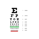
Visual Acuity Assessment
A non-invasive test that examines how well you can see up close and far away.
VISUAL ACUITY ASSESSMENT
In some instances, this is done for legal purposes such as determining if a patient is fit to drive a car. This 5-minute test is generally accomplished by looking at a chart containing letters of different sizes and reading the letters back to a technician.
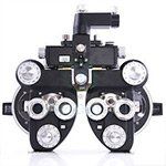
Refractions
An extension of a visual acuity test.
REFRACTIONS
This non-invasive test, performed manually or with a machine, determines the strength of the corrective lenses (glasses) or contacts that are needed for you to see clearly.
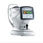
Wavefront Abberometry
Mapping the unique visual 'fingerprint' of how your eye bends light.
WAVEFRONT ABBEROMETRY
Using the same shack-hartmann technology utilized by NASA in its space telescopes, our wavefront aberrometer allows us to examine the unique properties of how your eye bends light. This information is then used for numerous purposes, from determining candidacy for LASIK or premium multifocal lenses for cataract surgery, to making highly accurate corrective lenses (glasses).
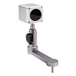
Meibomian Gland Imaging
A means of visualizing the meibomian glands, the glands that produce oil to keep your tears from evaporating away.
MEIBOMIAN GLAND IMAGING
Our eyes produce tears to lubricate the eye. However, in order for them to remain on the ocular surface, they require protection from evaporation. The Meibomian Glands, located in your lids, produce oil that prevent tear film evaporation. In some cases, the Meibomian Glands become clogged meaning oil cannot get to the ocular surface. Left untreated, these clogged oil glands will lead to permanent gland loss. Since these glands cannot be fully visualized by human eyes, we are proud to introduce this special camera to our office for the benefit of our patients for early detection of Meibomian Gland Dysfunction.
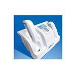
Tear Lab Tear Osmolarity Test
The tear osmolarity test is used to measure the composition of your eye’s tears.
TEAR LAB TEAR OSMOLARITY TEST
The results of this test give insights into whether the tears you produce are good enough to lubricate the cornea. Poor tears or not enough tears result in Dry Eye Disease. This test takes roughly 2 minutes to complete. During this time, a technician will obtain a small sample of your tears from the corner of your eye and analyze it in the Tearlab machine.
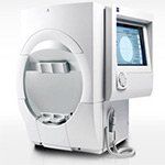
Visual Field (VF) Test
A non-invasive test that examines a patient's central and peripheral field of view.
VISUAL FIELD (VF) TEST
The results from this test notify the eye physician of any significant changes the patient has in their field of view. These data are very useful in the early diagnosis and monitoring of, glaucoma patients, macular degeneration patients, patients that have received trauma to the eye, as well as patients on certain drugs that may be toxic to the eye (e.g. plaquanil).
During the test, the patient will sit and stare at a fixed point within the analyzer. The analyzer will then flash a series of lights in various places while the patient clicks a button to indicate that he / she has seen the flash.
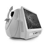
Optical Coherence Tomography (OCT)
A non-invasive test that utilizes light to examine the different layers of tissue in the retina.
OPTICAL COHERENCE TOMOGRAPHY (OCT)
By measuring the thickness of these tissues, eye physicians can better detect, diagnose, and treat certain retinal (or corneal) diseases, especially macular degeneration and diabetic retinopathy. It is also useful in assessing the health of the optic nerve, which may be damaged by diseases such as glaucoma.The test is generally conducted while dilated and takes a few minutes to complete. During that time, the patient sits and stares at a fixed point while the OCT scans the eye. The procedure is then repeated for the other eye.
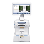
Endothelial Cell Count
A means by which the number of corneal endothelial cells are quantified.
ENDOTHELIAL CELL COUNT
We are all born with a fixed number of corneal endothelial cells which naturally die over time and are not replaced. However, trauma and certain eye diseases, such as glaucoma and uveitis, can accelerate the loss of these cells, potentially necessitating a corneal transplant. By quantifying these cells, eye physicians can track the status of the endothelium and be better able to detect and treat ailments before irreversible damage occurs.
This non-invasive test takes roughly 3 minutes to complete. During this time, the patient will sit down and stare at a fixed point while the cell counter images the eye via a flash photo. The procedure is then repeated for the other eye.
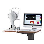
Anterior Segment Tomography
High Resolution, 3-D reconstruction of the front of the eye.
ANTERIOR SEGMENT TOMOGRAPHY
A non-invasive means by which a 3-D reconstruction of the front of the eye is be generated and analyzed for diseases of the front of the eye, such as Keratoconus. Results from this test can also be used to examine the thickness of the cornea, needed for accurate Tonometry, as well as examine refractive errors, such as astigmatism, with high resolution for specialized glasses, contact lens fits or LASIK evaluations.
This test takes roughly 5 minutes to complete. During this time, the patient will sit down and stare at a red/blue dot while a camera rotates around the eye.
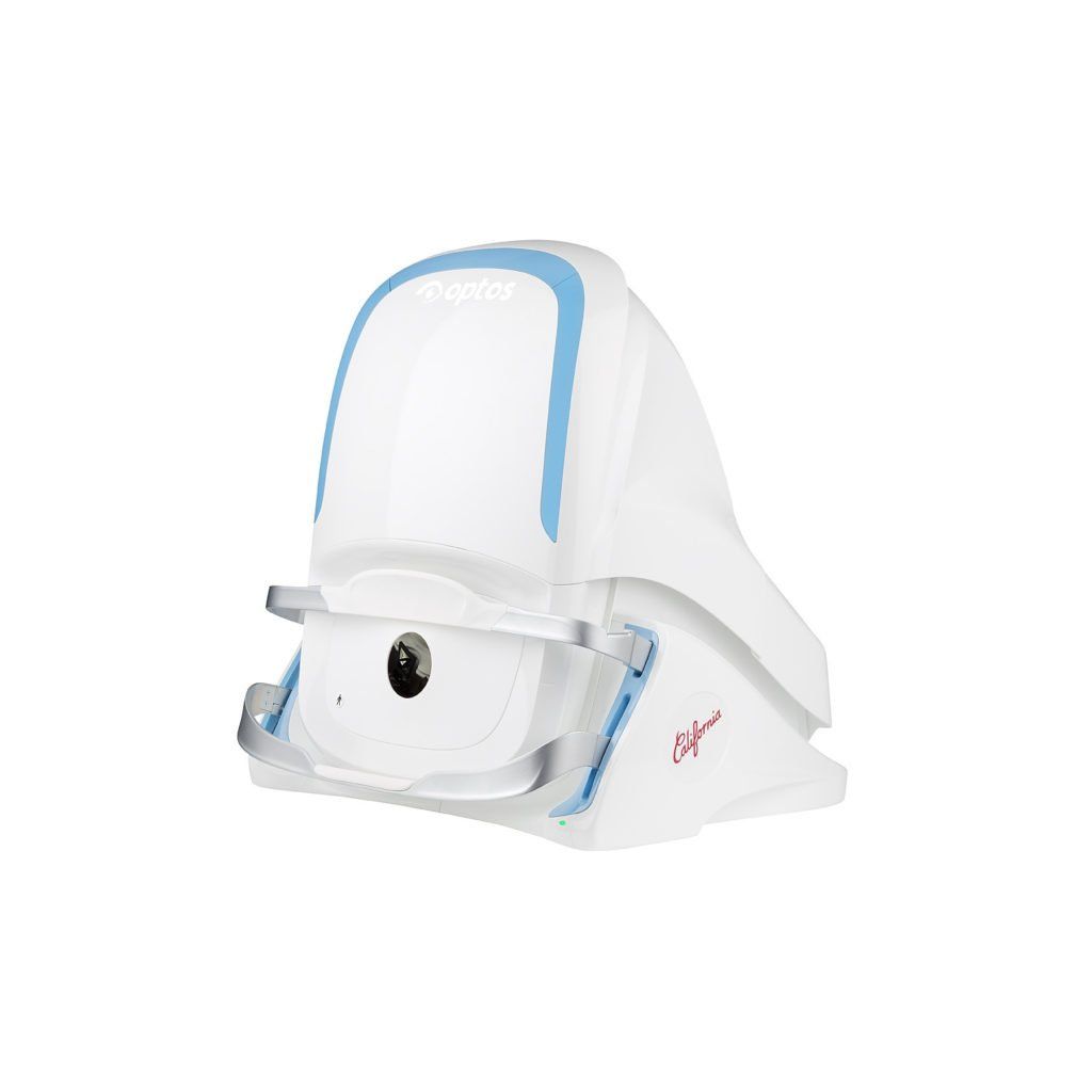
Ultra-WideField Fundus Photography
A non-invasive way to image large portions of the retina for future comparison.
ULTRA-WIDEFIELD FUNDUS PHOTOGRAPHY
While most cameras can only image 45 degrees of the retina, our confocal scanning laser ophthalmoscope can image 200 degrees, allowing our physicians to see into the periphery of the retina where pathology can go unnoticed. Our camera can also perform auto-fluorescence imaging to examine damage to the retina as well as fluorescein angiography to visualize blood vessels.
This test takes 5 minutes to complete. For this test, the patient will sit in front of the camera and view a target that will change color when the eye is properly focused. A flash of light will be then seen during image capture.
Non-Contact Comprehensive Biometry
A touch-free method to assess four different parameters that are critical for good cataract surgery outcomes.
NON-CONTACT COMPREHENSIVE BIOMETRY
These include measuring how long the eye is, how physically big the eye is, what the curvature of your cornea is, and how deep the area from your iris to your cornea is. This data, measured to the hundredth of a millimeter (or one tenth of a thousandth of an inch), is then statistically analyzed against advanced mathematical formulas that help determine the strength of your lens implant. Measurements taken here can allow most patients to enjoy freedom from glasses after their cataract surgery, should the patient desire.
This test takes roughly 5 minutes to complete. During this time, patients will be asked to fixate on a light while the device analyzes all four parameters. The test is then repeated on the other eye.
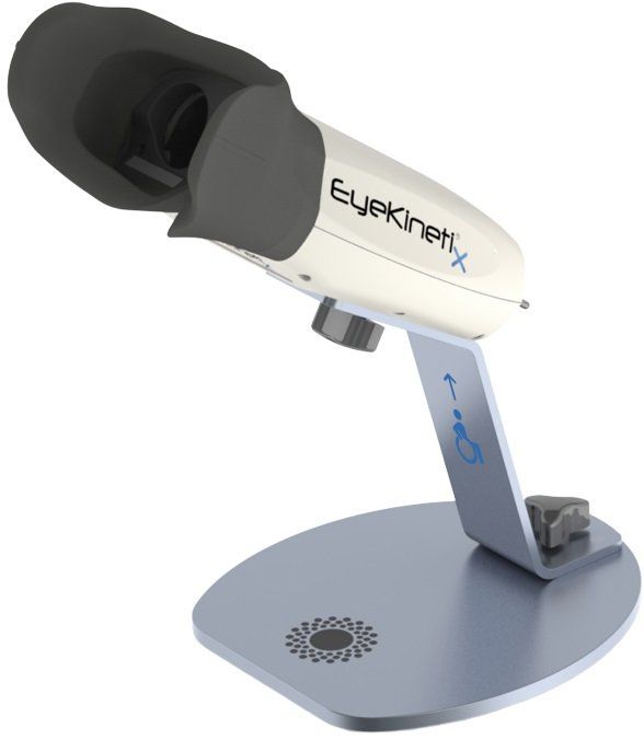
Quantitative Pupillometry
A highly reproducable, objective, and quantitiative approach to screening for afferent pupillary defects, or APDS, caused by asymmetric retinal or optic nerve disease.
Quantitative Pupillometry
The classic way of screening for afferent pupillary defects, or APDs, involves holding a penlight to one eye and watching the pupil response in the other. When one eye responds to light differently, it can be a sign of damage to the retina / optic nerve. This test, when performed manually, is difficult to accurately perform, relatively insensitive, and is highly variable between observers. The introduction of quantitative pupillometry, a first in the Capital Region, allows us to perform a much more sensitive and objective evaluation.
This test takes roughly 1 minute to complete. During this time, patients will be asked to fixate on a target while the device flashes light at one eye at a time. The test is then repeated on the other eye.
CONTACT INFO
3140 Troy-Schenectady Rd
Latham, NY 12309
Office Phone:
518-782-7777
HOURS
Office: Mon - Fri: 8:00 AM - 5:00 PM
1st Wed, every month: 9:00 AM - 5 PM
On call 24/7 for current patients
Optical: Mon - Fri: 8:30 AM - 5 PM
1st Wed, every month: 9AM - 5 PM
Optician Hours: 8:30 AM - 11:30AM;
12:30 PM - 5 PM
Eye physicians Schenectady NY, Diabetic Eye Doctor Albany NY, Lasik Eye Surgery, Glaucoma Doctors , Eye Exams In Saratoga, Schenectady, and Albany NY
Copyright 2024, Dr. Mabel Cheng, MD, PLLC| All Rights Reserved | *Content featured on this site is not designed to self-diagnose